On behalf of the Organizing Committee of Structural Biology 2019, all the researchers, developers, and experts are warmly welcome from the field of Structural Biology to attend 16th International Conference on Structural Biology schedule on March 11-12, 2018 in Bali, Indonesia. We aim to unify all the people indulged in this vast field and share the knowledge, explore and look forward to the new way by integrating new thoughts and customizing the limits of the future technology.
Structural Biology 2019 offers a dais to take an attention to explore, grip and meet with prominent speakers of the field, including both broad and specific subjects. The Structural Biology Meet 2019 will be surrounding the theme “Exploring recent trends in Structural Biology”.
The Structural Biology 2019 constitutes Sessions like Keynote Speeches, Oral Presentations, Poster Presentations, Universal Workshops, B2B Meetings, Panel Discussions, Q&A sessions, Industry expert interactions. There are awards for some categories like Best Poster, Best Oral presentation, Young Researcher Forums (YRF), e-Poster presentations, Video presentations by the experts from both Industry & Academic.
International Conference on Structural Biology 2019 invites all interested participants to join us for this venerate event at the elegant destination of Bali, Indonesia.
WHY TO ATTEND?
International Conference on Structural Biology is amid the World's leading technical Congress. The two days event on Structural Biology will host 60+ Scientific and technical sessions and sub-sessions on leading and latest research transformation in the field of Structural Biology covering the globe. Structural Biology 2019 will comprise of 20 major sessions designed to offer comprehensive sessions that discourse current topics in the field of Structural Biology.
The attendees can find exclusive sessions and panel discussions on the latest innovations in Structural Biology by:
-
Lectures from renowned speakers
-
Keynote forums by Prominent Professors, Engineers
-
Open Innovation Challenges
-
Poster presentations by Young Researchers
-
Global Networking sessions with 50+ Countries
-
Novel techniques to benefit your research
-
Best platform for Global business and Networking opportunities
-
Meet the editors of refereed journals, Society and Association members across the Globe
-
Excellent platform to showcase the latest innovation and concept in the technical field
Target Audience:-
-
Biochemists
-
Molecular Diagnosticians
-
Biotechnologists
-
Pharmaceutical Leaders
-
Cell Biologists
-
Developmental Biologists
-
Micro Biologists
-
Geneticists
-
Molecular Biologists
-
System Biologists
-
Immunologists
-
Biophysicists
-
Structural biologists
-
Nanoengineers
-
Neuro-Biologists
-
Bioengineers
-
Computational Biologist
-
Biomedical Researchers
-
Biomedical and Biotechnology Research organizations
-
Structural Biology Societies and Associations
-
Molecular Biology Societies and Associations
-
Professors & Academicians
-
Industrialist
-
Students
Track 1: Structural Biology
Structural biology is a branch of molecular biology, biochemistry, and biophysics involved with the molecular structure of biological macromolecules, how they acquire the structures they have, and how alterations in their structures affect their function. For a long time, we have possessed the capacity to imagine structures inside a cell, however, even the most ground-breaking magnifying instruments are constrained in the detail they give, either by the absolute physical limits of amplification or in light of the fact that the examples themselves are not alive and working. Auxiliary science methods dig underneath these breaking points breathing life into atoms in 3D and into more honed core interest. It ranges to the plain furthest reaches of how a particle functions and how its capacity can be altered.
Track 2: Proteomics and Genomics
Proteomics includes the study of composition, structure, function, and interaction of the proteins directing the activities of each living cell. It is the branch of molecular biology concerned with the study of the proteome.
Genomics is the sub-discipline of genetics devoted to mapping, sequencing and functional analysis of genomics. It studies all the genes present in an organism. It includes the study of intragenomic phenomena such as heterosis, epistasis, pleiotropy and other interactions between loci and alleles within the genomes.
Biochemistry is the branch of science in which deals with the study of the chemical and physical processes that occur in an organism. It integrates an introduction to the structure of macromolecules and a biochemical approach to cellular function. It includes the study of things as the structures and physical properties of biological molecules, including proteins, carbohydrates, lipids, and nucleic acids; the mechanisms of enzyme action; the chemical regulation of metabolism; the chemistry of nutrition; the molecular basis of genetics; the chemistry of vitamins; energy utilization in the cell; and the chemistry of the immune response.
Biophysics involves the study of biological systems and biological processes using physics-based methods or based on physical principles. It is an interdisciplinary research area between physics and biology. Biological phenomena and Living nature can be explored using the principles and methods applied in physics.
-
Industrial & Structural Biochemistry
-
Clinical & Forensic Biochemistry
-
Pharmaceutical & Metabolic Biochemistry
-
Biochemistry in Cancer Treatment Research
-
Animal & Plant Biochemistry
-
Food & Nutritional Biochemistry
-
Biophysical Techniques
-
Biophysical approaches to cell biology
-
Nanobiophysics
-
Biophysical Mechanisms to explain specific biological processes
Bioinformatics is an interdisciplinary field that develops method and software tools for understanding biological data. It combines various fields including computer science, statistics, mathematics, and engineering to analyze and interpret biological data. It is a hybrid science that links biological data with techniques for information storage, distribution, and analysis to support multiple areas of scientific research, including biomedicine. It is fed by high-throughput data-generating experiments, including genomic sequence determinations and measurements of gene expression patterns.
Molecular Biology is the field of biology that studies the composition, structure, and interactions of cellular molecules like nucleic acids and proteins which carry out the biological processes essential for the cell's functions and maintenance. It covers a wide scope of problems that are related to molecular and cell biology including structural and functional genomics, transcriptomics, proteomics, bioinformatics, biomedicine, molecular enzymology, molecular virology, and molecular immunology, etc.
Microbiology involves the study of the micro-organisms that are too small to be seen by naked eyes. It includes eukaryotes such as fungi and protists and prokaryotes such as bacteria and certain algae.
Enzymology involves the study of enzymes, their kinetics, structure, and function, as well as their relation to each other. Enzymes play an important role in all metabolic and cellular signaling pathways. Scientists are often interested in understanding how enzymes function at the molecular and atomic level through a combination of modern biochemistry and structural biology. Techniques used to investigate enzyme structure and dynamics include X-ray crystallography, NMR, mass spectroscopy and protein chemistry, while their chemical behavior is being characterized by rapid-reaction and steady-state kinetics, calorimetry, chemical analyses, and a variety of spectroscopies. Protein engineering is also used to study how structure determines function.
Synthetic Biology is an interdisciplinary branch, combining the principles of biology and engineering in order to produce biological functions in organisms that may or may not otherwise occur naturally. It is an emerging field that aims at designing and building novel biological systems.
Viruses use a molecular mechanism to penetrate the host cells to confirm an infection and to assure that the progeny virus particles are discharged into the environment invading the host’s immune defenses. This phenomenon is known as Structural Virology. Although viruses are simple as the individual with the self-replicating ability but as a group, they are exceptionally diverse in strategies and structures.
3-D structures determination of proteins is crucial for the understanding of these interactions as well as their structure-function relationships, which also has many practical applications in drug design and protein engineering. Determining the 3D structure of protein help us to understand the mechanism of action of protein and its functions.
Computational approaches are benefits for structural biology. Structure of molecules is determined by experimental methods which are tedious and practical. Computational biology is an interdisciplinary field that develops and applies computational methods to analyze large collections of biological data, such as genetic sequences, cell populations or protein samples, to make new predictions or discover new biology. The computational methods used include analytical methods, mathematical modeling, and simulation. It is a rapidly developing multi-disciplinary field. The systematic achievements of data made possible by genomics & proteomics technologies have created a tremendous gap between available data & their biological interpretation.
-
Ab Initio Method
-
Threading method
-
Homology modeling
Molecular modeling is a computer-based technique for determining and representing the three-dimensional structures of the molecules, also the properties associated with these 3-D structures. The main applications of molecular modeling are the generation of protein structures, molecular structure visualization, and determination of molecular properties.
Molecular dynamics simulation is a technique to study the structure and functional relationship of macromolecules. It mimics the real-life motion of atoms and molecules.
-
Potentials In Molecular Dynamics
-
Steered Molecular Dynamics
-
Molecular Dynamics Algorithms
-
Incorporating Molecular Dynamics
-
Design Constraints
Structural bioinformatics is a special practical result for protein structure determination. Structural Bioinformatics is an interdisciplinary field that deals with the three-dimensional structures of biomolecules. It attempts to model and discover the basic principles underlying biological machinery at the molecular level. It is based on the hypothesis that 3D structural information of a biological system is the basic to understanding its mechanism of action and function. Structural bioinformatics combines applications of physical and chemical principles with algorithms from computational science. Major areas protein and nucleic acid 3D structure determination, prediction of protein 3D structure from sequence, protein structure validation protein structure comparison and alignment, protein and nucleic acid structure classification inferring protein function from structure, prediction of protein-ligand interaction, prediction of protein-protein interactions, development of databases.
-
Hybrid of experimental methods
-
NMR structures
-
Hybrid of computational methods
-
Determining protein complex structures
-
Bottom-up integration of atomic detail crystallography
-
Hybrid approaches in complementing high-resolution structural biology
Structural biologist aims to determine the structure of the protein and along with drug designing such as the identification of hits, leads, and candidate drugs. Protein plays an essential role in all biochemical reactions that occur in the body. They act as carriers and providing strength and structure. Discovering the structure of a protein has always been tedious and complex. Innovative ideas are being improved in different fields of structural biology.
The main part of research is being carried out in the area of cancer. The principal aim is to design and discover innovative and efficient drugs to cure the disease. Structural biology combined with molecular modeling principally aims at drug designing. Ultimately, many researchers in Structural biology carry out cancer research to increase the exploitation of molecular understanding of biomolecules in the advancement of innovative cancer therapies. Cancer immunotherapy is also used which is defined as the response of the immune system to reject cancer. Immunotherapies can be a beneficial way of treating cancer. The malignant tumor cells are attacked by stimulating the immune system as these cells are capable to exploit the fact that cancer cells often have molecules on their surface that can be detected by the tumor-associated antigens (TAAs).
A biological macromolecule's function is known by the chemical and physical properties of its threeâ€dimensional (3D) structure. Concerning this, one should know the structure of a biomolecule which is very important if we want to know the living systems and diseases. A database is a structured combination of data. In the field of structural biology immense research is being done and as the result, huge data is being produced. To store the data in an organized manner, bioinformatics databases are used. Many databases are designed so that the biological data can be stored such as sequence databases, signaling database, structure database, etc. The Protein database(PDB) which is a crystallographic database is used for 3D structural data of larger biomolecules. The advancement in technologies had been considered in the further development of the PDB and in the structural specialty and structural characteristic databases that have also evolved. The mainly used databases are Electron Microscopy Data Bank, Protein Structure Classification Database (CATH), Structural Classification of Protein (SCOP) and Protein Data Bank (PDB).
-
Classification of structural database
-
Classification of protein structure
-
Protein structure classification database
-
Protein data bank
-
Electron microscopy data bank
Structural biology is one of the developing fields. In the course of time, many modifications have taken place. A large number of resolved structures have increased rapidly. The drug design and drug discovery field have been advanced. Another field where progressions are being seen is functional annotations. Modifications, in order to improve the effectiveness of prevailing tools, can also be remarked. Extraordinary advances can be seen in the areas of imaging technologies and the advancement of hybrid methods to explain the structure and function of proteins. Structural biology is one of the progressing fields. In the course of time, many developments have been taking place.
-
Structure Determination
-
Technological Advances In Existing Methods
-
New And Potentially Disruptive Technologies
-
Advances In Drug Design
-
Advances In Tool Development
-
Advances In Imaging Technologies
Drug design is an inventive process to find new medication centered on the knowledge of the biological target. It is also known as also known as rational drug design. A drug is most a small molecule that inhibits or activates the function of a biomolecule, which in results into a therapeutic benefit to the patient. Drug design commonly relies on computational techniques. This type of modeling is often mentioned as a computer-aided drug design. Drug design that depends on the knowledge of the 3D structure of the target is known as structure-based drug design. The main methods available for drug design are structure-based drug design and ligand-based drug design. The structure-based drug designing also known as direct drug design involves the three-dimensional structure of a drug target interacting with small molecules is used to guide drug discovery.
Biomarkers are biological measures of a biological state. It is a characteristic that is accurately measured and evaluated as an indicator of natural biological processes, pathogenic processes or pharmacological responses to a therapeutic intervention.
-
Drug designing
-
Drug development
-
Drug modelling
-
Drug targeting
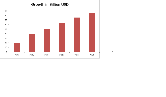
Glance at Market of Structural Biology
The concerned region of Structural Biology Analyzer industry market involves North American, Europe, and Asia etc., and the main countries include United States, Germany, Japan, and China etc. North America is the leading region in Structural Biology Analyzers following the European market which is the second largest market for Structural Biology Analyzers. The constant healthcare sector improvements and huge population base represented by the Asia Pacific region is expected to drive the importance in Asia Pacific Biochemistry & Structural Biology Analyzers market.
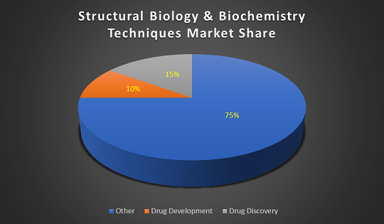
The Structural Biology Analyzers market offers a healthy contribution to the In-Vitro Diagnostic market and is expected to grow in the upcoming years. The global market for in vitro diagnostics (IVD) products was $60.3 billion in 2015 and expected to be $81.1 billion by 2020 at a compound annual growth rate (CAGR) of 6.1%.
North America leads the global IVD products market throughout the period, worth $24.6 billion in 2014. The market is expected to reach $29.4 billion in 2020 from $25.3 billion in 2015 increasing at a CAGR of 3.1%. Asia is the fastest growing region of the global IVD market with a CAGR of 12.9% from 2015 to 2020. The market is worth $15.3 billion in 2015 and is expected to reach $28.2 billion by 2020.
Why Bali, Indonesia?
Bali is a province of Indonesia and an island on the westernmost of the Lesser Sunda Islands, located on the east of Java and west of Lombok. The province includes the island of Bali and a few smaller neighboring islands, notably Nusa Penida, Nusa Lembongan, and Nusa Ceningan. Bali is the only Hindu-majority province in Indonesia, with 83.5% of the population adhering to Balinese Hinduism. Bali is Indonesia's main tourist destination, which has seen a significant rise in tourists. It is renowned for its highly developed arts, including traditional and modern dance, sculpture, painting, leather, metalworking, and music. Indonesia's most popular vacation spot Bali’s ticks all the boxes for an exotic destination, with endless ribbons of the beach, traditional villages, unspoiled jungles and forests, and ornate ancient temples.
Major Structural Biology Related Associations around the Globe:
-
American Society for Biochemistry and Molecular Biology
-
Czech Society for Structural Biology
-
Indonesian Protein Society
-
Asia Pacific Federation for Clinical Biochemistry and Laboratory Medicine
-
Italian Proteomics Association
-
Proteomics Society, India
-
Biophysical Society
-
American Chemical Society
-
New England Structural Biology Association
-
The Protein Society
-
American Crystallographic Association
-
International Union of Crystallography
-
British Crystallographic Association
-
European Crystallographic Association
-
Swiss Proteomics Society
-
Hellenic Crystallographic Association
-
Bioinformatics society of India
-
International Society for Computational Biology
-
South African Crystallographic Society
-
Australian Society for Biochemistry and Molecular Biology
-
Australian Society for Biophysics
-
American Society for Mass Spectrometry
-
British Biophysical Society
-
Bioinformatics Italian Society
-
Mid-South Computational Biology and Bioinformatics Society
Major Structural Biology Related Research Units :
-
Structural Biology Group, RIKEN
-
OKINAWA Institute of Science and Technology
-
Markey Center for Structural Biology
-
New York Structural Biology Center
-
Department of Anatomy and Structural Biology, Einstein College
-
Department of Structural and Cellular Biology - Tulane University
-
Structural Biology — Penn State University
-
Chemical and Structural Biology – The Rockefeller University
-
Center for Structural Biology - University of Illinois at Chicago
-
Structural Biology Facility - Robert H. Lurie Comprehensive Cancer Center of Northwestern University
-
Department of Cellular and Structural Biology – UT Health Science Center
-
UCSF Macromolecular Structure Group
-
Structural Biology NMR Facility - University of Minnesota
Related Conferences
-
13th International Conference on Structural and Molecular Biology: Techniques & Market Analysis, October 22-23, 2018 Ottawa | Ontario | Canada
-
World Structural and Molecular Biology Conference, November 26-28, 2018, Rome, Italy
-
World Congress on Advanced Structural and Molecular Biology, February 27-28, 2019, Paris, France
-
11th Edition of International Conference on Structural Biology March 07-08, 2019, Berlin, Germany
-
15th World Congress on Structural Biology, March 18-19,2019, PARIS, FRANCE
-
15th International Conference on STRUCTURAL AND MOLECULAR BIOLOGY, March 20-21, 2019 Sydney, Australia
-
4th International Conference on Biochemistry & Molecular Biology, May 10-11, 2019, TOKYO, JAPAN
-
2nd International Conference on Computational Biology and Bioinformatics, May 17-18, 2019, SINGAPORE CITY, SINGAPORE
-
21st International Conference on Structural Biology, June 17 - 18, 2019, Toronto, Canada
-
18th International Conference on Structural Biology, October 14-16, 2019 London, UK
New method exposes structures inside “rainbow†of brain cells
Molecules from alpacas may enable scientists to identify cell types in the brain while also revealing their interior structures. The method may help researchers better visualize how the brain is wired in autism. Researchers often stain tissue with fluorescent antibodies to certain proteins so they can identify cell types. They can also use electron microscopy to view the tissue’s internal structure.
But the two techniques can’t be used on the same sample. The former requires chemicals that often break down cells. Electron microscopy involves thinly slicing the sample, and staining the numerous slices afterwards is prohibitively laborious. Scientists have engineered brain cells to produce fluorescent proteins or made tags that are visible in an electron microscope. But these methods can label only about three cell types per sample. The new technique lets researchers tag tissue with up to 10 markers.
The researchers stained sections of mouse brain using fluorescent ‘nanobodies’ antibody fragments typically derived from camelids, the mammalian family that includes camels, alpacas and llamas. Because of their small size, nanobodies can infiltrate tissue without it needing to be pretreated with harsh chemicals. The team created images of the stained tissue with a fluorescent microscope. They then sliced the samples into 50-nanometer-thick segments and scanned those with an electron microscope. Using computer software, they aligned both sets of images based on landmarks such as blood vessels.
The resulting images highlight four cell types, including star-shaped support cells called astrocytes and immune cells called microglia, each labeled with different colors. The fluorescent nanobodies penetrate more than 100 micrometers into the block of tissue. By contrast, antibody markers stain only the surface. The images show organelles such as mitochondria and microtubule bundles. The researchers could also make out the long filaments that extend from neurons, and the vesicles at the end of them that hold chemical messengers. The nanobody method could reveal differences in cell structure and the distribution of cell types in the autism brain.
The ultimate goal is to stain a sample with hundreds of colors by developing more nanobodies and combining fluorescent dyes.
Taking a closer look
Cryo-electron microscopy, an emerging technology that can show the structure of molecules down to the atomic level with more clarity than ever before. Years ago, we were able to see only the general areas where the proteins were as blobs on a screen but now it is possible to see the atoms within those blobs. This could help researchers design new drugs that better target diseases or create new compounds that speed up or slow down chemical reactions.
Electron microscopes use a beam of electrons instead of rays of light to create an image. Because electrons have a much shorter wavelength than visible light, such microscopes can provide better resolution of molecules at the atomic level. What we are looking at is a projection of how the electrons interact with the sample. Since everything is made of electrons, they repel each other. Thus, what is generating is a sort of two-dimensional projected image of your sample. Cryo-EM is a modification of the standard electron microscope that involves freezing a sample in solution so rapidly that the surrounding water forms a thin, glass-like layer instead of crystallizing. Scientists can then take multiple images of that frozen sample and piece them together to create a remarkably clear 3-D image.
Seeing the shape and arrangement of the molecule could help scientists better understand how these molecules work. Some proteins operate very much like tiny machines, using a chemical process to create mechanical energy. They have very mechanical properties, so when we see the whole thing, then we immediately start to see what the pieces are and how they move. With cryo-EM scientists can take various images of these proteins and put them together like a stop-action movie to see how those mechanical properties work.
As technology has improved it’s opened up new areas of research. Pharmaceutical companies, for example, are using cryo-EM to map out the sites where drugs can bind to the target cells, so they can better identify which drugs might affect those cells and learn what part of the protein they interact with.
Tiny straws improve molecule delivery
Researchers have found what they believe may be a better way to deliver molecules directly into human cells they require to manipulate. “Nanostraws” are small glass-like protrusions that poke equally little holes in cell walls before releasing their cargo that is completed more safely and with efficiency than existing strategies.
They say breakthrough might speed up medical and biological research and in the future will improve gene therapy for cancer and diseases of the eyes and immune systems. Scientist expertise with nanomaterials was the key to the new approach, that improves on the method referred to as electroporation, whereby an electrical current is employed to form holes in cell walls.
Existing electroporation strategies will be imprecise, usually killing several of the cells researchers try to work with. Nanostraws are more economical as a result of their long, narrow profile helps concentrate electrical currents into a really tiny space.
They’re additionally a far better possibility than approaches based mostly around using viruses or alternative chemicals to carry molecules across cell walls. The researchers initially tested their technique on animal cells sitting atop a bed of nanostraws. when they turned on the electrical current, the nanostraws opened little, regularly sized pores within the cell membrane: enough to permit molecules in, however not enough to try to serious injury. the current drew molecules straight into the cell, further increasing the speed and precision of the method.
The question at that time was whether or not the technique would be as effective on the kinds of human cells clinicians would wish to manipulate to treat diseases. They successfully delivered molecules into 3 human cell types as well as mouse brain cells, all of that had proven difficult to work within the past. The technique was fast and killed fewer than 100% of cells, an enormous improvement on normal electroporation, they say.
The next step is to test it with human immune cells, that are among the hardest to figure with.
The study reveals how a small molecule promotes the removal of excess cholesterol
Experts have discovered the structure of the activated form of an enzyme that helps to return excess cholesterol to the liver.
The analysis reveals how a drug-like chemical stimulates the action of the lecithin: cholesterol acyltransferase (LCAT) enzyme. It also recommends that future drugs using the same mechanism could be used to restore LCAT function in people with familial LCAT deficiency (FLD), a rare inherited disease that settles them at risk of eye problems, anemia, and kidney failure.
LCAT helps high-density lipoprotein (HDL) known as the good cholesterol to remove cholesterol from the blood by converting the lipid into a form that is accessible to package and transport. There are more than 90 known mutations in LCAT, which can cause either a partial loss of activity (known as 'fish-eye disease') or full loss (FLD). Boosting LCAT activity may be beneficial in treating people with coronary heart disease and LCAT deficiencies, but the mechanisms by which it can be activated are poorly understood.
In this study, structural biology is used to understand how a patented LCAT activator binds to LCAT and how it promotes cholesterol transport. It was also studied if the compound could help recover the activity of LCAT enzymes that have commonly observed mutations seen in FLD.
The researchers used X-ray crystallography to look at the LCAT enzyme stabilized in its active state with two different chemicals, the activator molecule, and a second compound that mimics a substrate bound to the enzyme. The two chemicals had more of an effect on the protein when presented together than when presented separately, which suggested that they bind to the enzyme in different places.
Further analysis has found that the activator molecule, unlike other known LCAT activators, binds to a region close to where HDL attaches. However, the activator did not help LCAT bind to the HDL more efficiently, which led the researchers to speculate that it instead helps to transfer cholesterol and lipids into the catalytic center of the enzyme, so that it can convert it into cargo for transport in HDL.
On establishing this mode of action, the researchers tested whether this molecule could help recover the cholesterol-transport function of a mutant LCAT enzyme. They made a version of the enzyme with a mutation commonly seen in FLD patients and then tested its ability to bind to HDL and convert cholesterol in the presence or absence of the activator molecule. They were delighted to find that the activator could partly reverse the loss of activity in the mutant enzymes, resulting in comparable cholesterol conversion to the normal enzyme.
The results will help scientists to design compounds that can better target LCAT so that they might be of therapeutic benefit for heart disease and FLD patients. Future efforts will be to examine whether patients with other LCAT genetic mutations could benefit from the compounds used in this study and to design molecules with improved pharmacological properties for further development.
Structural study of antibiotic opens the way for new TB treatments
A new analysis of the structure and function of the naturally-occurring antimicrobial agent tunicamycin has shown ways to produce new, safe antibiotics for Mycobacterium tuberculosis and other disease-causing bacteria.
Tunicamycin is an antimicrobial produced by different types of bacteria, but it is inappropriate for use in humans because it is also toxic to animal cells.
In a new investigation, researchers studied the mechanism behind Tunicamycin's toxicity and pronounced that it acted upon a gene called DPAGT1, which is capable of producing an enzyme required in glycoprotein biosynthesis. This is a gene that often mutates, leading to rare genetic diseases.
By analyzing the interaction of Tunicamycin with this protein the researchers were able to know how it acts upon cells. The researchers also altered its structure to create new analogs of the antibiotic, several of which were useful in treating Tuberculosis (TB) in mice, while also being less toxic than alternative medicine presently wont to treat the malady.
TB poses a growing threat as our arsenal of drugs to treat it is rapidly working out. An exciting outcome from this research work is the development of a promising new class of antimicrobial drug.
This research represents a really beautiful synergy of the striking structural biology of a key human enzyme and the redesign of one of the most famous natural products in science, now redesigned to form a brand new family of antibiotics for TB.
Tuberculosis is a disease caused by the bacteria Mycobacterium tuberculosis, which causes around 1.3 million deaths each year globally. The huge majority of deaths occur in emerging countries where poor sanitation and access to healthcare compound the problem. Antibiotic resistance is turning into a major drawback with TB as several strains of the disease have become resistant to a variety of the drugs available, and a few strains resistant to all of them.
In the future, being able to design new drugs in a way that ensures their lower toxicity compared with rivals represents a real game-changer in the search for new treatments.